Hemifusomes: A Newly Described Organelle in Human Cells Implicated in Vesicle Biogenesis and Cellular Recycling
Author: Jonathon James Vukani Wigley | Nd. Nat. Con (1997); MEd (2006) | SkillsLink Academy (Pty) Ltd
Abstract
A previously unrecognized organelle, the hemifusome, has been discovered in mammalian cells. Identified by in situ cryo‑electron tomography, hemifusomes are heterotypic vesicle complexes connected by an extended hemifusion diaphragm and associated with proteolipid nanodroplets. Comprising up to 10% of peripheral vesicles, these structures define an ESCRT‑independent pathway for multivesicular body formation and cellular recycling. This paper reviews the morphology, distribution, putative function, and potential disease relevance of hemifusomes, and discusses future research directions in cellular physiology and genetic disorder investigation.
Introduction
Intracellular trafficking and cargo sorting via vesicles and multivesicular bodies (MVBs) are essential for cell homeostasis. Current models rely heavily on ESCRT‑mediated inward budding for MVB formation. Despite decades of research, predicted intermediate membrane states such as hemifusion diaphragms have eluded in situ visualization in live cells (Tavakoli et al., 2025). Recent employment of cryo‑electron tomography (cryo‑ET) has now revealed the presence of a novel organelle, the hemifusome, that challenges existing paradigms of vesicle biogenesis (Tavakoli et al., 2025) PMC+14Nature+14IFLScience+14.
Methods (Summarized)

Researchers used cryo‑ET to image four mammalian cell lines—COS‑7, HeLa, RAT‑1, and NIH/3T3—preserved in a near-native frozen state to minimize processing artifacts (Tavakoli et al., 2025) Nature+1Earth.com+1. Peripheral cellular regions of 150–400 nm thickness were imaged at ~300 kV, enabling visualization of membrane-bound vesicles including the hemifusomes Nature.
Results
Morphology and Prevalence
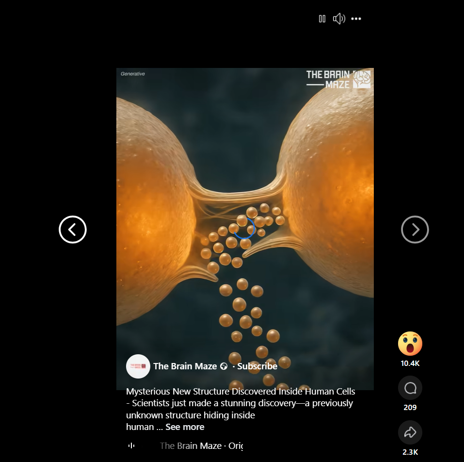
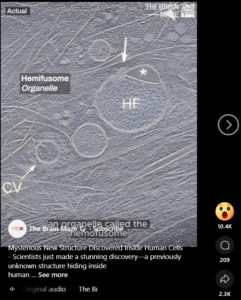
Hemifusomes appear as two heterotypic vesicles sharing an expanded hemifusion diaphragm (≈160 nm), often with a ~42 nm proteolipid nanodroplet (PND) anchored at the rim UVA Health Newsroom+13Nature+13Wikipedia+13. These structures represented up to ~10 % of vesicular organelles at the cell periphery Nature+1Wikipedia+1.
Structural Configurations
Two main conformations were identified:
-
Direct hemifusomes: a smaller and a larger vesicle share the hemifusion diaphragm.
-
Flipped hemifusomes: an intraluminal vesicle hemifused to the luminal side of a larger vesicle Sci.News: Breaking Science News+4Nature+4Earth.com+4.
Both types are consistently associated with PNDs, and compound (multi-vesicle) hemifusomes were also observed Technology Networks+11Nature+11Earth.com+11.
Functional Implications
Unlike canonical endocytic pathways, hemifusomes do not engage ESCRT-mediated mechanics. Rather, they seem to offer a lipid‑based remodeling alternative for MVB formation via PND‑mediated vesicle biogenesis Earth.com+4Nature+4Wikipedia+4. They are thus proposed as loading‑dock–like platforms for intracellular cargo sorting and membrane recycling Live Science+10UVA Health Newsroom+10SciTechDaily+10.
Discussion
Cellular Role and Mechanism
The hemifusome may stage an intermediate membrane state long hypothesized in models of vesicle fusion and scission (hemifusion diaphragm), but now directly observed in situ Wikipedia+5Nature+5Earth.com+5. This suggests a vesicle formation pathway distinct from ESCRT, potentially driven by membrane physics and lipid droplets rather than protein scaffolding.
Disease Relevance
Defects in cargo handling—particularly in multivesicular body formation—are implicated in conditions such as Hermansky–Pudlak syndrome, which affects pigmentation, lung function, vision, and coagulation Medicine in Motion NewsThe Sun+5UVA Health Newsroom+5Earth.com+5. The malfunction of hemifusomes may represent a previously unrecognized pathogenic mechanism across various genetic and neurodegenerative diseases Drug Target ReviewTechnology NetworksScienceAlert.
Future Directions
Key next steps include mapping hemifusome prevalence across tissues and cell types, determining their life cycle and regulation, identifying associated proteins and lipidochemical composition, and probing their behavior in disease models. Experimental perturbation (e.g. PND inhibitors, knockdowns) could test causal links to cargo trafficking and pathophysiology.
Conclusion
The hemifusome represents a transformative discovery in cell biology: a structurally and functionally distinct organelle involved in vesicle biogenesis and material recycling. Its consistent presence, unique topology, and potential disease connections open novel investigative paths. This finding underscores the continuing importance of high-resolution, native-state imaging in uncovering the hidden architecture of living cells.
References
Tavakoli, A., Hu, S., Ebrahim, S., & Kachar, B. (2025). Hemifusomes and interacting proteolipid nanodroplets mediate multi‑vesicular body formation. Nature Communications, 16(1), 4609. https://doi.org/10.1038/s41467‑025‑59887‑9 Live Science+14Nature+14PMC+14Wikipedia+11Medicine in Motion News+11The Sun+11
University of Virginia Health System. (2025, June 25). Scientists discover unknown organelle inside our cells. UVA Health Newsroom. Sci.News: Breaking Science News+11UVA Health Newsroom+11SciTechDaily+11
Technology Networks. (2025, June 25). New organelle discovered with roles in cellular recycling. Technology Networks. Technology Networks
ScienceAlert. (2025, July 14). Mysterious new structure discovered hiding inside human cells. ScienceAlert. ScienceAlert
Drug Target Review. (2025, July 3). Meet the hemifusome: a new organelle with big impact. Drug Target Review. Drug Target Review
Live Science. (2025, June 30). Scientists discover never‑before‑seen part of human cells — and it looks like a snowman wearing a scarf. Live Science. Wikipedia+2Live Science+2The Sun+2
Sci.News. (2025). Hemifusomes: Biologists discover previously unknown organelle. Sci.News. Sci.News: Breaking Science News


